[Complete Collection] Drug target model of CD74-ROS1 + DNA standard + RNA standard + ddPCR kit
ROS proto-oncogene 1 (ROS1), encoded by the ROS1 gene located on chromosome 6q22.1, belongs to the insulin receptor subfamily of tyrosine kinases and was discovered in 1986 in a study involving the sarcoma RNA UR2 tumor virus. The physiological role of ROS1 remains controversial; its expression was observed mainly in lung tissue, followed by cervix and colon. Conceivably, the tyrosine kinase insulin receptor plays a key role in embryonic development. The ROS1 protein shows substantial homology with ALK (both belonging to the insulin receptor superfamily), especially in the ATP-binding site (84% homology) and the kinase domain (64% homology). Fusion mutations in ROS1 are well known and often result in gene fusions with several fusion partners, and the resulting fusion proteins are powerful oncogenic drivers. Thus, ROS1 kinase activity is persistently activated, resulting in increased cell proliferation, survival, and migration due to upregulation of JAK/STAT, PI3K/AKT, and MAPK/ERK signaling pathways. ROS1 has shown tumorigenic potential in vitro and in vivo, and glioblastoma was the first human cancer shown to harbor ROS1 rearrangements. ROS1 fusions have subsequently been observed in other malignancies, including NSCLC, melanoma, and occasionally cholangiocarcinoma, angiosarcoma, ovarian, gastric, and colorectal cancers. ROS1 alterations are neither inherited nor acquired as genetic alterations.
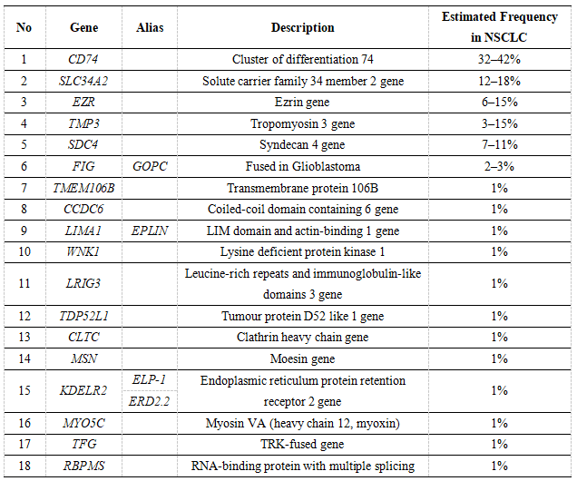
The rearrangement of ROS1 accounts for about 2% of NSCLC patients. The common fusion partners in NSCLC are shown in the table below, of which the highest proportion is the CD74 gene.

Table 1. Main ROS1 fusion partners in non-small cell lung cancer (NSCLC).
CD74 is located on chromosome 5q33.1, and the protein encoded by this gene is associated with the major histocompatibility complex (MHC) class II and is an important molecular chaperone that regulates antigen presentation in the immune response. It also acts as a cell surface receptor for the cytokine macrophage migration inhibitory factor (MIF), which when bound to the encoded protein initiates survival pathways and cell proliferation. This protein also interacts with amyloid precursor protein (APP) and inhibits the production of amyloid beta (Abeta).
In the diagnosis of non-small cell lung cancer, the NCCN guidelines clearly describe "ROS1 is a receptor tyrosine kinase that can be rearranged in NSCLC, resulting in dysregulation and inappropriate signaling through the ROS1 kinase domain." A molecular marker that must be detected, and the recommended detection method is described as "FISH break-apart probe methodology can be deployed; however, it may under-detect the FIG-ROS1 variant. IHC approaches can be deployed; however, IHC for ROS1 fusions has low specificity, and follow-up confirmatory testing is a necessary component of utilizing ROS1 IHC as a screening modality. Numerous NGS methodologies can detect ROS1 fusions, although DNA-based NGS may under-detect ROS1 fusions. Targeted real-time PCR assays are utilized in some settings, although they are unlikely to detect fusions with novel partners.”
One of the Complete Collections: The Drug Target Model
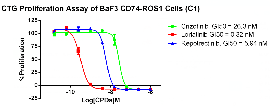
CD74-ROS1 fusion protein can constitutively activate cell proliferation. Selected on the IL3-dependent BaF3 cell model, exogenous overexpression of CD74-ROS1 protein can be used as a driver gene to drive the proliferation of Baf3 cells without IL3, so it can For activity detection of inhibitors against CD74-ROS1. Based on the above principles, we have developed a CD74-ROS1/Baf3 drug target model, which can detect the inhibitory activity of samples at the in vitro level, and can also make a CDX model at the in vivo level to detect the activity of the drug.

Fig 1. Sanger sequencing of CD74-ROS1/BaF3(CBP73331), CD74 gene on the left and ROS1 gene on the right.

Fig 2. Cell-based kinase assay of CD74-ROS1/BaF3(CBP73331), comparison of the inhibitory activities of different positive drugs on the proliferation of CD47-ROS1 cells.
The Complete Collection Part 2: DNA Standards
We know that CD74 is located on chromosome 5, and ROS1 is located on chromosome 6. The CD74-ROS1 fusion is a translocation fusion. The common CD74(E6)-ROS1(E34) is in the DNA sequencing of clinical patients. The breakpoint of CD74 It is on intron 6, the breakpoint of ROS1 is on intron 33, and different cases, the breakpoint is not unique, so based on the standard of CD74-ROS1 at the DNA level, first of all, the breakpoint must be On introns, a second standard represents only one breakpoint. Based on the above information, we developed and customized a DNA-level CD74(E6)-ROS1(E34) through gene editing technology, calibrated the DNA through sanger sequencing and ddPCR detection, and released a DNA standard.

Fig 3. Sanger sequencing of CD74-ROS1 Translocation (CBP20177D), CD74 gene on the left, ROS1 gene on the right, and the breakpoints are intron breakpoints.

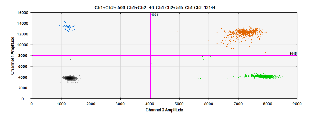
Fig 4. ddPCR of CD74-ROS1 Translocation (CBP20177D), the detection showed that the mutation frequency of this standard was 50%.
Complete Collection No. 3: RNA Standards
CD74-ROS1 fusion is pathologically meaningful, mainly because of the activity of the fusion protein. That is to say, assuming that fusion occurs at the DNA level, but there is no RNA, and thus no protein, it does not make any sense. Therefore, the RNA targeting CD74-ROS1 detection would be more meaningful. Through gene recombination technology, we customized the cell model of CD74(E6)-ROS1(E34) at the RNA level, and then calibrated the RNA by sanger sequencing and ddPCR detection, and released an RNA standard.

Fig 5. Sanger sequencing of CD74-ROS1 Fusion (CBP20170R), CD74 gene on the left, ROS1 gene on the right, and the breakpoints are exon breakpoints.

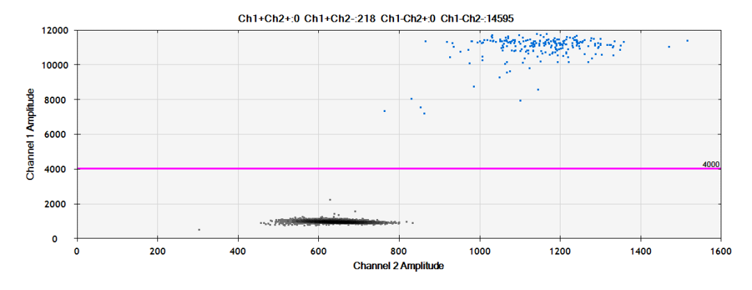
Fig 6. DdPCR of CD74-ROS1 Fusion (CBP20170R), the test showed that the copy number of this standard was 2244.5 copies/ng.
Complete Collection Four: ddPCR Kit
Whether it is drug development or diagnosis for CD74-ROS1 fusion, PCR detection technology is indispensable in various application detections. For example, in the construction of drug target models, we can use PCR technology to check whether exogenous genes are overexpressed. , look at the positive ratio, screen clones, methodological optimization, etc. For example, in diagnosis, positive patients can be screened, performance evaluation, usual quality control, etc. In PCR technology, ddPCR is a method of absolute quantification with high sensitivity. Compared with Q-PCR, it has high sensitivity and does not require a standard curve. Compared with NGS, it has lower cost and shorter cycle, and can express results. We have developed ddPCR detection kits for DNA and RNA, suitable for detection in drug development and clinical diagnosis.

Fig 7. The DNA-level ddPCR performance evaluation for CD74-ROS1, using the standard diluted to different %AF, showed accurate detection ability at different %AF.

Fig 8. ddPCR performance evaluation at the RNA level for CD74-ROS1, using standard dilutions to different copy numbers, showed accurate detection ability at different copy numbers (note that RNA needs to be reverse transcribed into cDNA for detection, reverse transcription requires carried out under the exact same system).

